Home

visuel Travaux ménagers amical candida albicans mikroskop auxiliaire Inciter Serpent

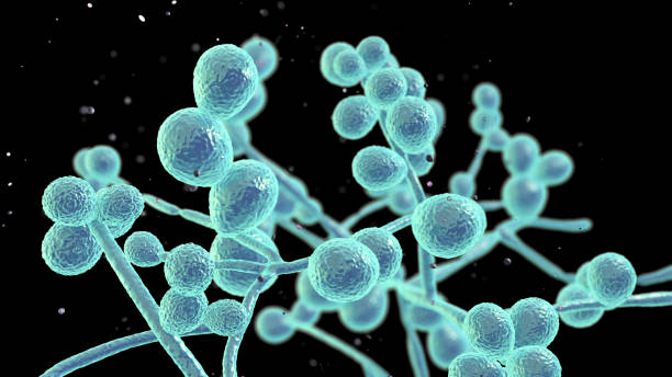
Bildagentur | mauritius images | Color enhanced scanning electron  micrograph (SEM) of the yeast-like fungus Candida albicans - a cause of  thrush (candidiasis). This fungus is normally present on human skin and
Bildagentur | mauritius images | Color enhanced scanning electron micrograph (SEM) of the yeast-like fungus Candida albicans - a cause of thrush (candidiasis). This fungus is normally present on human skin and

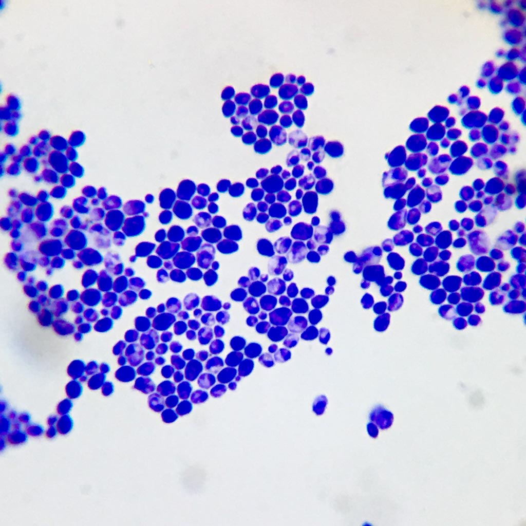
Candida Albicans Colonies Under Microscope Stock Photo 1558658411 |  Shutterstock
Candida Albicans Colonies Under Microscope Stock Photo 1558658411 | Shutterstock

Microscopic View Of Candida Albicans From A Patient With Mycosis 19th  Century Stock Illustration - Download Image Now - iStock
Microscopic View Of Candida Albicans From A Patient With Mycosis 19th Century Stock Illustration - Download Image Now - iStock

Candida Albicans I Mikroskop Fotografering för Bildbyråer - Bild av agape,  prövkopia: 177868269
Candida Albicans I Mikroskop Fotografering för Bildbyråer - Bild av agape, prövkopia: 177868269

Candida albicans (Pathogenesis) - microbewiki

34 Blastospore Images, Stock Photos & Vectors | Shutterstock

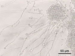
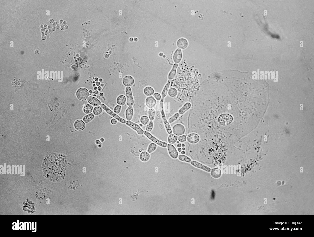
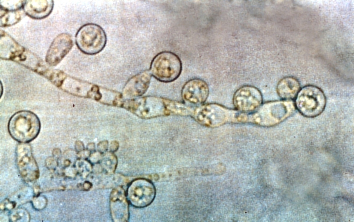
File:Candida albicans (optic microscope).jpg - Wikimedia Commons

Candida albicans – Wikipedia

Microscopic feature of yeasts (100x). A : Candida albicans (A1 :... |  Download Scientific Diagram
Microscopic feature of yeasts (100x). A : Candida albicans (A1 :... | Download Scientific Diagram

Candida albicans – Wikipedia

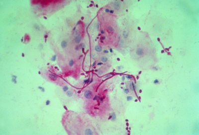
File:Candida albicans (sputum).jpg - Wikimedia Commons

Candida albicans was examined under oil immersion at 100× magnification...  | Download Scientific Diagram
Candida albicans was examined under oil immersion at 100× magnification... | Download Scientific Diagram

162: Candida albicans – Fungus Fact Friday

Candida albicans smear, Fungi prepared slides expert

Premium Photo | Candida colony under microscopic view or candida albicans  fungus infection
Premium Photo | Candida colony under microscopic view or candida albicans fungus infection

Candida albicans micrograph hi-res stock photography and images - Alamy

Candida Albicans And Bacterial Colonies Under The Microscope Stock Photo,  Picture and Royalty Free Image. Image 143837057.
Candida Albicans And Bacterial Colonies Under The Microscope Stock Photo, Picture and Royalty Free Image. Image 143837057.

candida albicans under microscope|TikTok Search

6,200+ Fungi Microscope Stock Photos, Pictures & Royalty-Free Images -  iStock
6,200+ Fungi Microscope Stock Photos, Pictures & Royalty-Free Images - iStock

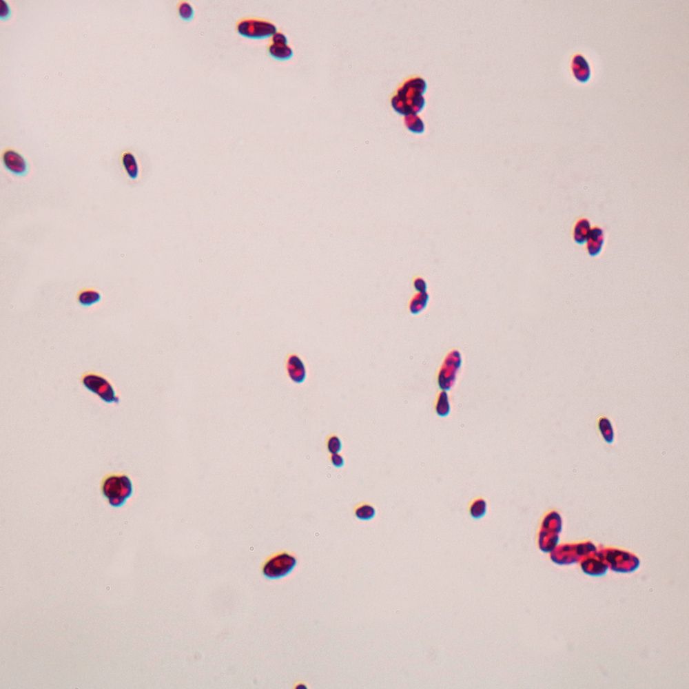
shows the shape the cell of Candida albicans ander the microscope |  Download Scientific Diagram
shows the shape the cell of Candida albicans ander the microscope | Download Scientific Diagram

Candida albicans - Wikipedia

Candida – Store norske leksikon

Candida Albicans Under The Microscope Stock Photo, Picture And Royalty Free  Image. Image 151639540.
Candida Albicans Under The Microscope Stock Photo, Picture And Royalty Free Image. Image 151639540.

Candida albicans, w.m. Microscope Slide | Carolina Biological Supply

Microscopic appearance of Candida albicans. A. Cells from stationary... |  Download Scientific Diagram
Microscopic appearance of Candida albicans. A. Cells from stationary... | Download Scientific Diagram